Scientists in Singapore and the US have developed a new method of detecting how scars will develop after a surgery or burn by using a breakthrough method with nanoparticles and a handheld fluorescent microscope.
This new method can help avoid biopsies and other invasive techniques.
Armed with this information, doctors can take preventative measures to avoid serious scarring.
This research was led by: “Assistant Professor Xu Chenjie from NTU’s School of Chemical and Biomedical Engineering, nanoscience expert Professor Chad A Mirkin from Northwestern University, United States, and Dr. Amy S Paller, Chair of Dermatology at Northwestern University Feinberg School of Medicine.” [1]


New Pain-Free Technique
As noted by the Nanyang Technological Institute, “Clinicians currently find it difficult to predict how scars will develop following surgery … without resorting to invasive testing.” [2] This new method can help avoid biopsies and other invasive techniques.
How does it work?
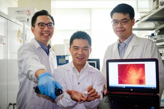
The article by ScienceDaily goes on to say “the new detection method uses thousands of nanoparticles called NanoFlares which have DNA strands attached to their surfaces like a ball of spikes.” [3] The nanoparticles are applied as a cream to the closed wound.
After the nanoparticles have penetrated the skin cells for 24 hours, a handheld fluorescence microscope (like the Dino-Lite shown) is used to look for signs of nanoparticles’ interaction with target biomarkers. If the microscope detects fluorescence signals, it indicates abnormal scarring activity. From there, doctors can take preventive action can hopefully avoid heavier scarring.