With the use of a variety of Dino-Lite fluorescence microscopes alone, we were able to identify the cells.
The AM4115T-GFBW is designed to observe green fluorescence including but not limited to green fluorescent protein (GFP). This model uses 1 White LED and 7 Blue LEDs at an excitation of 480nm and includes an emission filter at 510nm. Aside from the AM4115T-GFBW, we offer a variety of other fluorescence microscopes designed to observe specific proteins. For example, the AM4115T-RFYW is designed to observe red fluorescent protein (mCherry). This model uses 1 White LED and 7 Yellow LEDs at an excitation of 575nm and includes an emission filter at 610nm. Some of our models, like the AM4115T-GRFBY can even observe two different kinds of proteins. This model is designed to observe GFP and MCherry. This model uses 4 Blue LEDs at an excitation of 480nm and 4 Yellow LEDs at an excitation of 575nm which are switchable through the software.
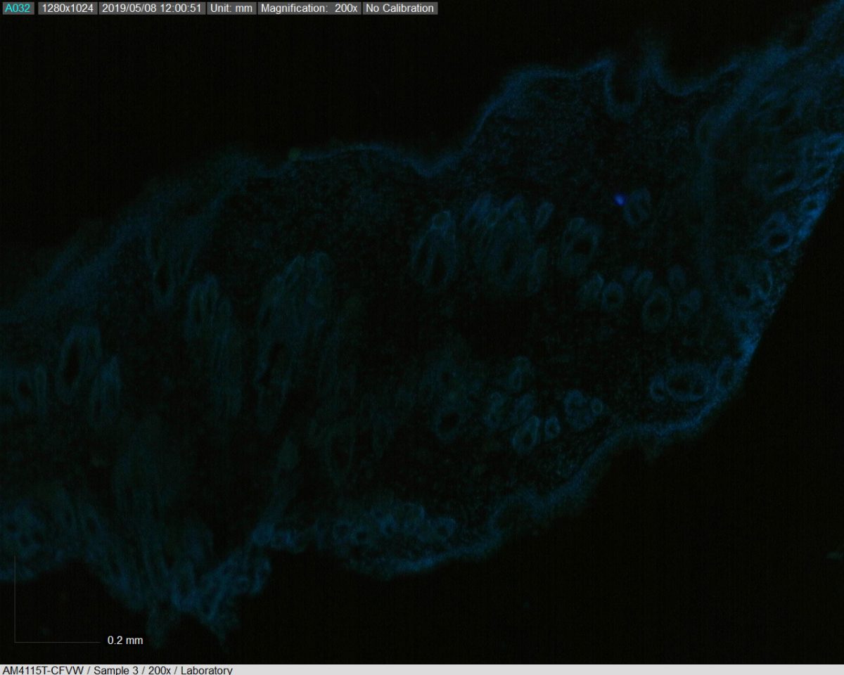
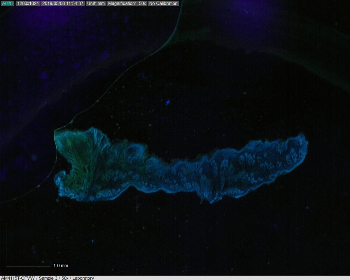
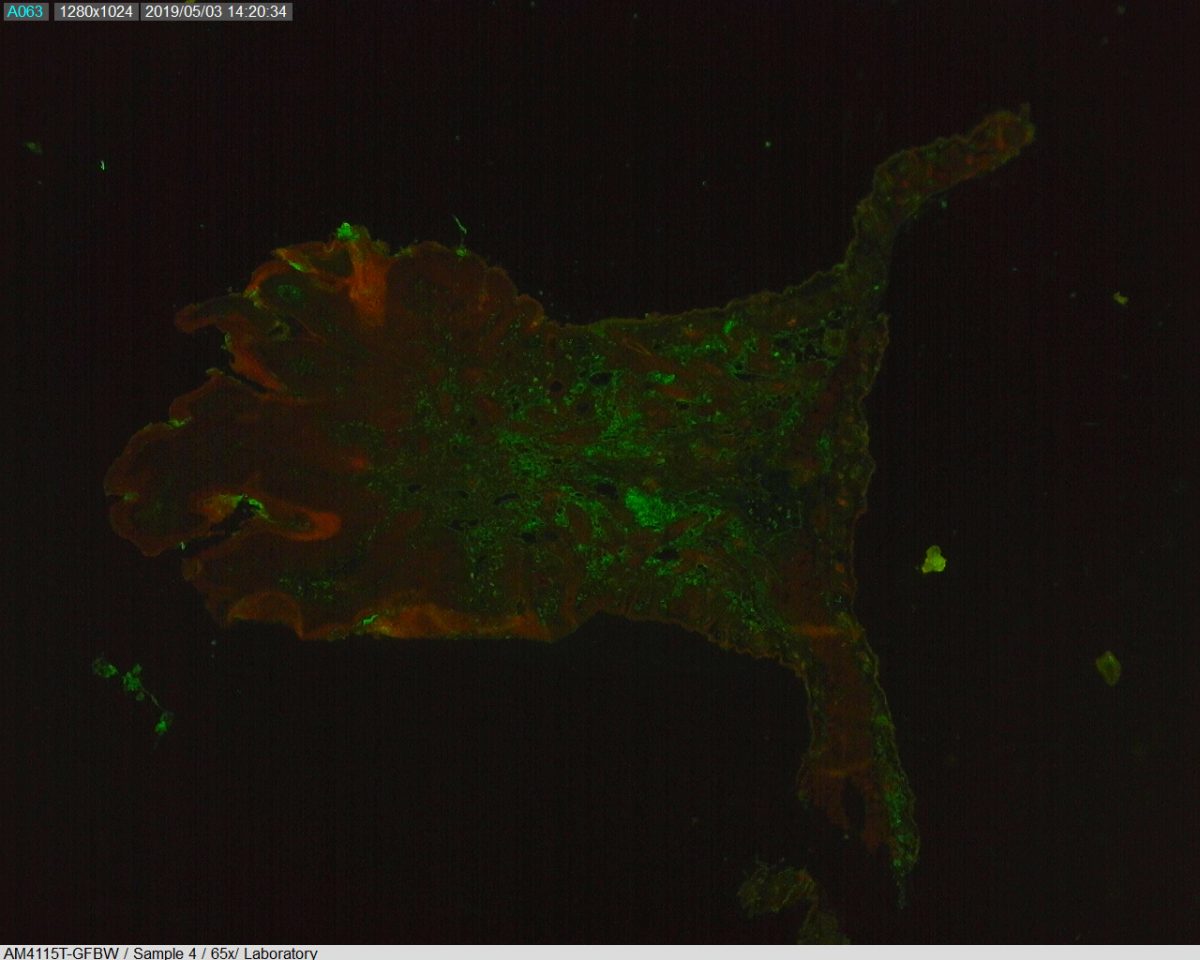
 Picture taken with: AM4115T-GFBW
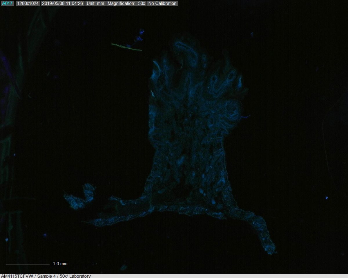
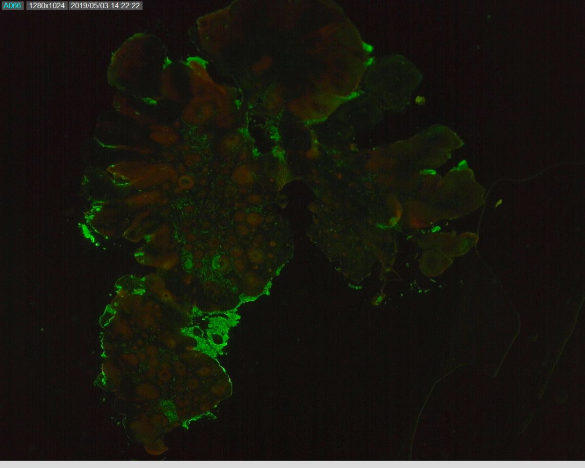
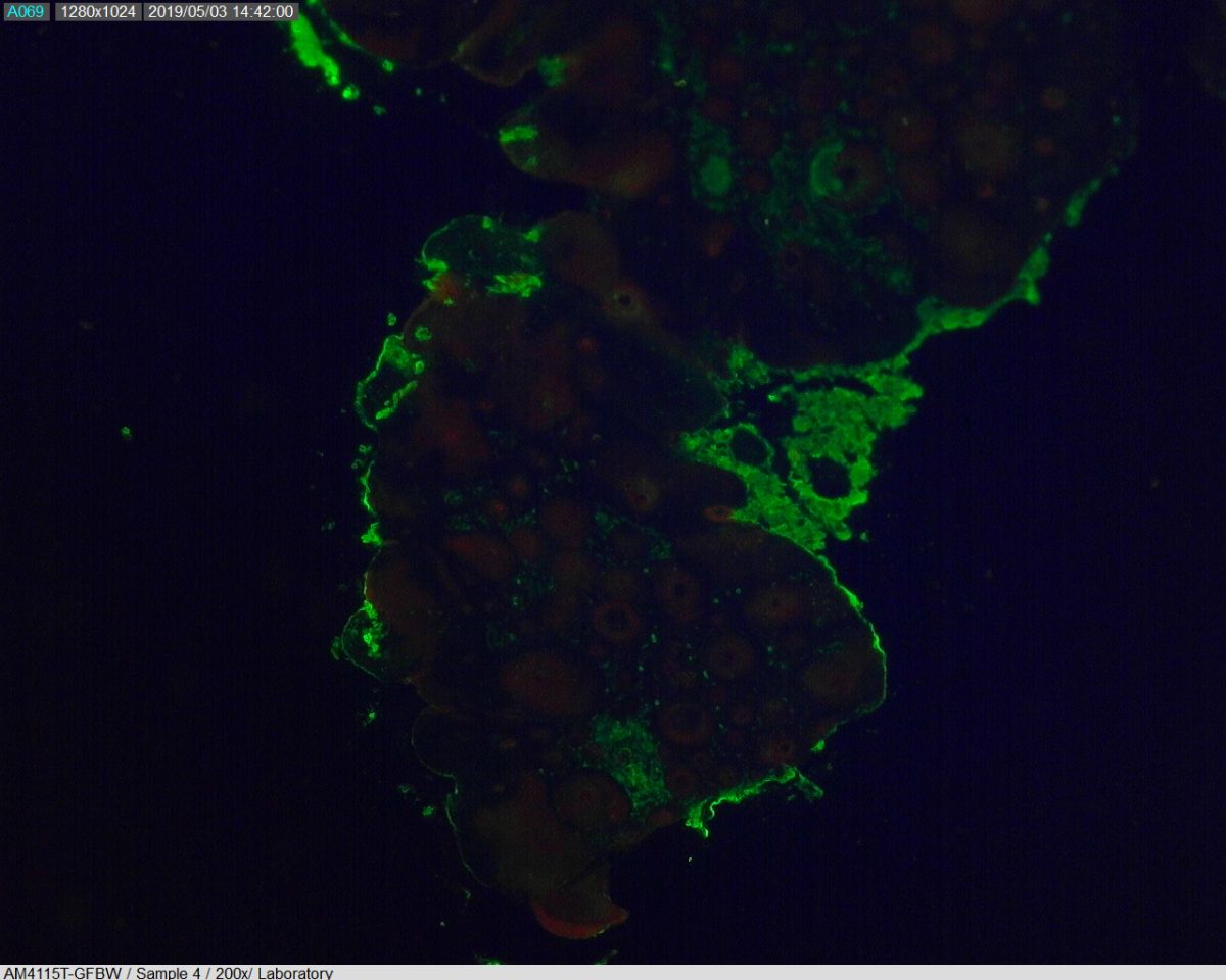
Slide details: Murine squamous cell carcinoma stained with Chicken Anti-Cytokeratin 5 and Rabbit Anti-Ki67. Secondary antibodies Goat Anti-Chicken AlexaFlour 488 (FITC) and Goat Anti-Rabbit AlexaFlour 594 (Texas Red). Mounted in ProLong Gold with DAPI nuclear counterstain.
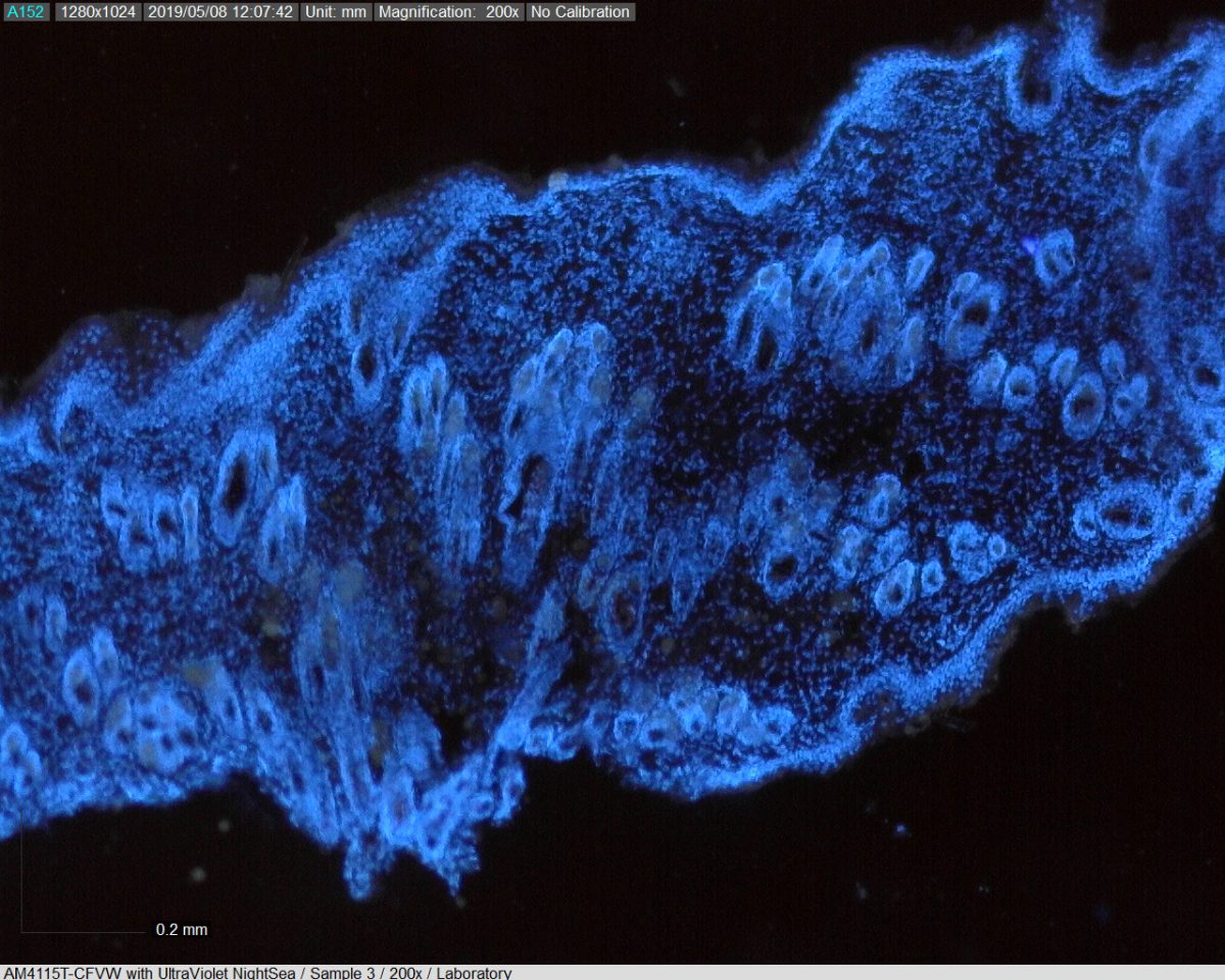
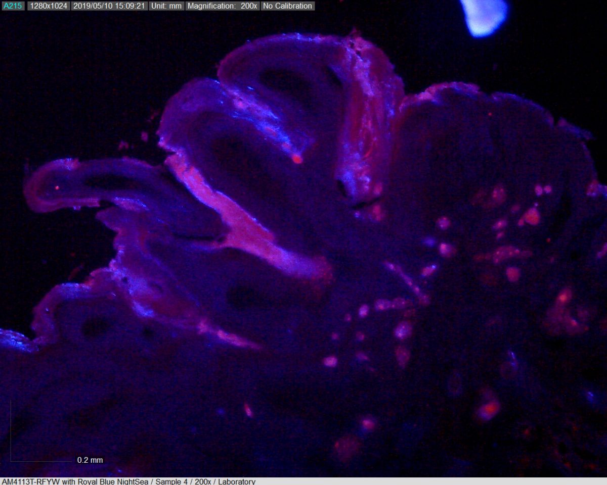
To increase the effectiveness of the built-in Dino-Lite LED’s while in high magnification, we took advantage of the premium offerings of NIGHTSEA; a company that provides light sources and matched filters for viewing and imaging fluorescence at scales from macro to micro.

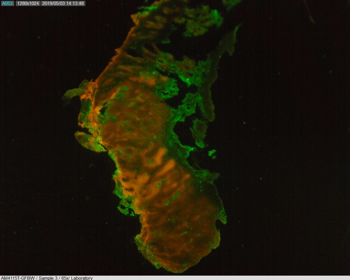
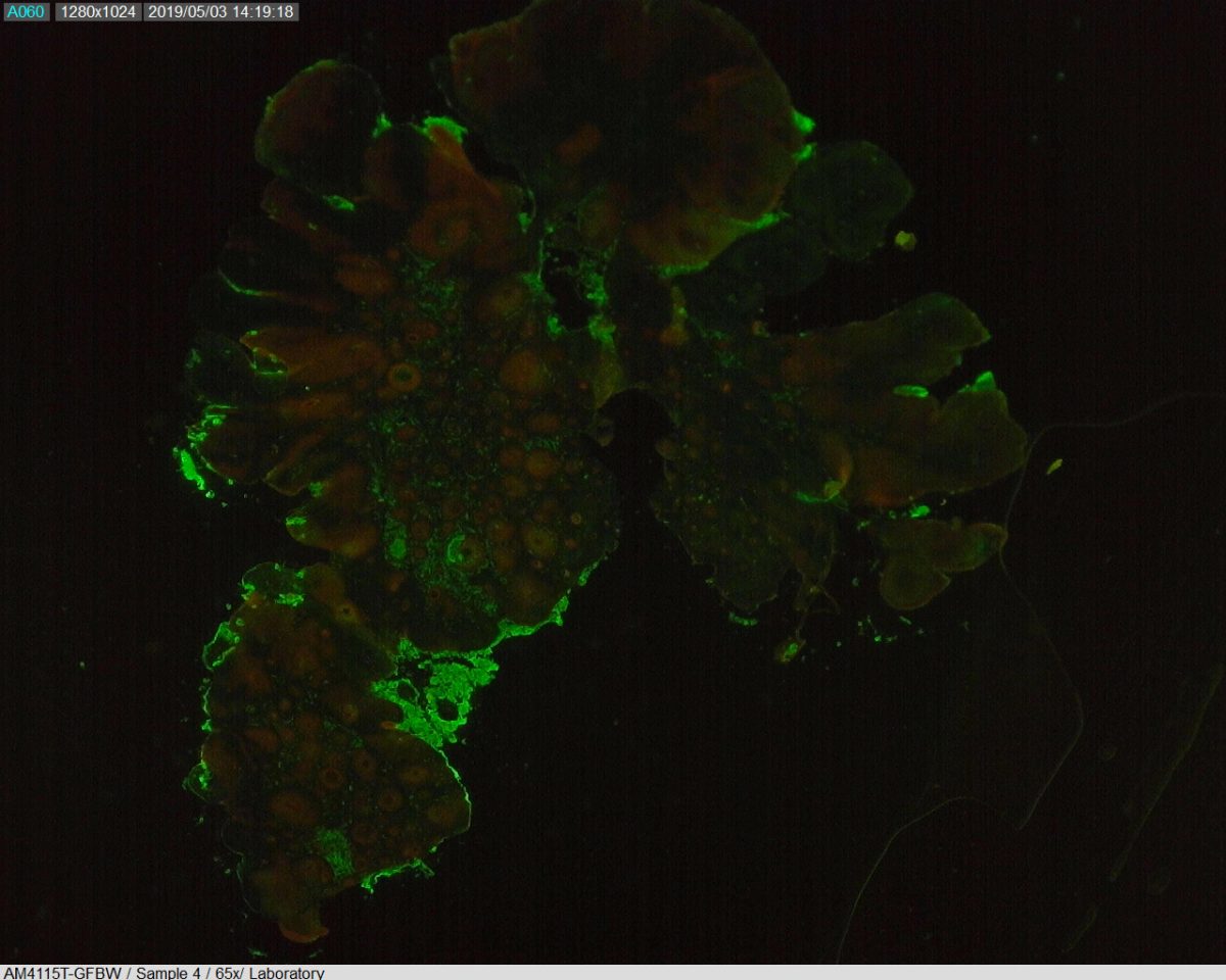
Picture taken with: AM7515MT8A-GFBW with NightSea’s Royal Blue excitation light.
Slide details: Murine squamous cell carcinoma stained with Chicken Anti-Cytokeratin 5 and Rabbit Anti-Ki67. Secondary antibodies Goat Anti-Chicken AlexaFlour 488 (FITC) and Goat Anti-Rabbit AlexaFlour 594 (Texas Red). Mounted in ProLong Gold with DAPI nuclear counterstain.
Fluorescence microscopy is a powerful tool that enables the study of diverse processes in the biomedical field. This versatility is the reason why more and more studies are being done using variants of fluorescent microscopy. Many new techniques have been developed in the past decade that enables for better imaging quality and Dino-Lite Fluorescence Models are part of that wave. In this simple testing, users were able to gather quality images of the stained cells with the Dino-Lite fluorescent microscope models along with NightSea. With a few adjustments, Dino-Lite microscopes appear to have quick and easy results as powerful as the traditional fluorescence microscopes.


